
Investigating soft fouling during lentiviral vector bacterial retention testing
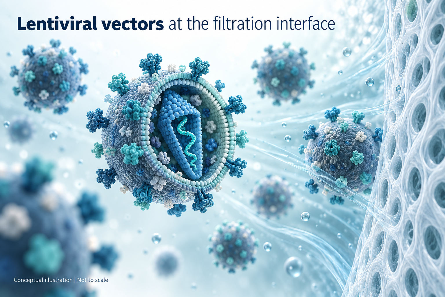
Figure 1 Conceptual representation of enveloped lentiviral vector particles near a membrane. The cutaway is illustrative; particle structure and relative dimensions are not represented quantitatively.
An unexpected pressure rise during bacterial retention validation can expose a difference between the behaviour of a lentiviral process stream and the same stream after addition of a concentrated bacterial challenge. Understanding that difference requires both operational evidence and an assessment of the underlying physical mechanisms.
An anonymised validation programme illustrates the issue. A vector-containing challenge showed substantially higher initial pressure and incomplete volume recovery, while a vector-free matrix study was recorded as valid. A subsequent written account reported altered particle size and concentration after mixing vector with the challenge organism. Together, these findings supported investigation of a challenge-associated interaction and soft fouling.
The practical task was to establish what the observations demonstrated, which explanations remained to be confirmed and how a scientifically justified retention study could be completed. In my view, filtration engineering, microbiology and vector analytics all have a role in that assessment.
The observed filtration behaviour
The programme included a vector-containing bacterial challenge using membrane discs and a separate challenge using the medium without vector in a larger capsule format. Each study comprised three runs and a positive control.[9]
The technical review described an approximately 20–40-fold difference in initial transmembrane pressure between the vector-containing and vector-free challenges. It also reported that the high-pressure behaviour was not observed in routine low-bioburden processing. These observations provided a substantive reason to investigate a challenge-associated restriction.
The comparison has an important limitation: filter format and area differed between the studies. It therefore does not isolate the effect of the vector or establish the size of that effect under matched conditions. The complete pressure, flow and loading records would be needed for that assessment.
The vector-containing disc study was classified as invalid. Organisms recovered downstream were identified as species other than the intended challenge organism, so their recovery cannot be interpreted as demonstrated breakthrough of that organism. The vector-free study provides evidence for the conditions it tested; it does not, by itself, validate the vector-containing process.
A further vector-containing capsule study was listed without reported results. Proposed follow-up work should therefore remain clearly distinguished from completed evidence.
| Study condition | Volume recovered | Microbiological result and study status |
|---|---|---|
| Vector-containing medium with bacterial challenge Membrane disc format | 20–39 mL from a 40 mL challenge | Reported counts 0, 0 and 14 CFU Recovered species differed from the challenge organism Classified as invalid |
| Medium without vector with bacterial challenge Larger capsule format | 40 mL from 40 mL in each of three runs | Reported counts 0, 0 and 0 CFU Classified as valid |
Anonymised summary of reported study observations. Dates, protocol numbers, product codes and organisation names have been removed. The conditions were not a matched filter-format comparison.
A lentiviral intermediate is a complex particulate system
Lentiviral vectors are enveloped particles whose filtration behaviour depends on more than their nominal size. Their surface properties, aggregation state, formulation and the accompanying process impurities can influence recovery and the development of hydraulic resistance.
Producer cells also release extracellular vesicles. Experimental work has demonstrated their presence in lentiviral preparations and the difficulty of separating particles with overlapping biophysical properties.[1] The residual profile after purification is process dependent; neither anion exchange chromatography nor tangential flow filtration should be assumed to remove every relevant colloid.
Consequently, a clear-looking intermediate may still contain material capable of interacting with a membrane. The investigation should establish the composition and condition of the actual feed rather than infer filterability from appearance alone.
The bacterial challenge changes the filtration environment
Brevundimonas diminuta is commonly used in bacterial retention studies for sterilising-grade filters. FDA guidance generally recommends a challenge of at least 10 million organisms per square centimetre of effective filtration area, providing a safety margin beyond the microbial burden expected in production.[2] This is a deliberate validation challenge, rather than a simulation of routine bioburden.
Adding that challenge introduces a substantial additional particulate population. The inoculum vehicle may also change the fluid composition. These differences can affect deposition and resistance, even without any specific association between bacteria and vector particles.
Possible physical association deserves consideration. A subsequent three-condition Videodrop experiment was reported to show a marked change in the measured particle population after bacteria and vector were mixed.[11] This strengthens the case for a challenge-associated interaction, but does not identify the components of the altered population or demonstrate direct binding. Controlled imaging or another method capable of distinguishing the components would help establish the mechanism.
This discussion concerns physical behaviour during filtration. It does not imply that lentiviral vectors infect the bacteria. It is also inappropriate to treat bacteria as universally rigid particles: published filtration research found pressure-dependent deformability in B. diminuta under the conditions examined.[3]
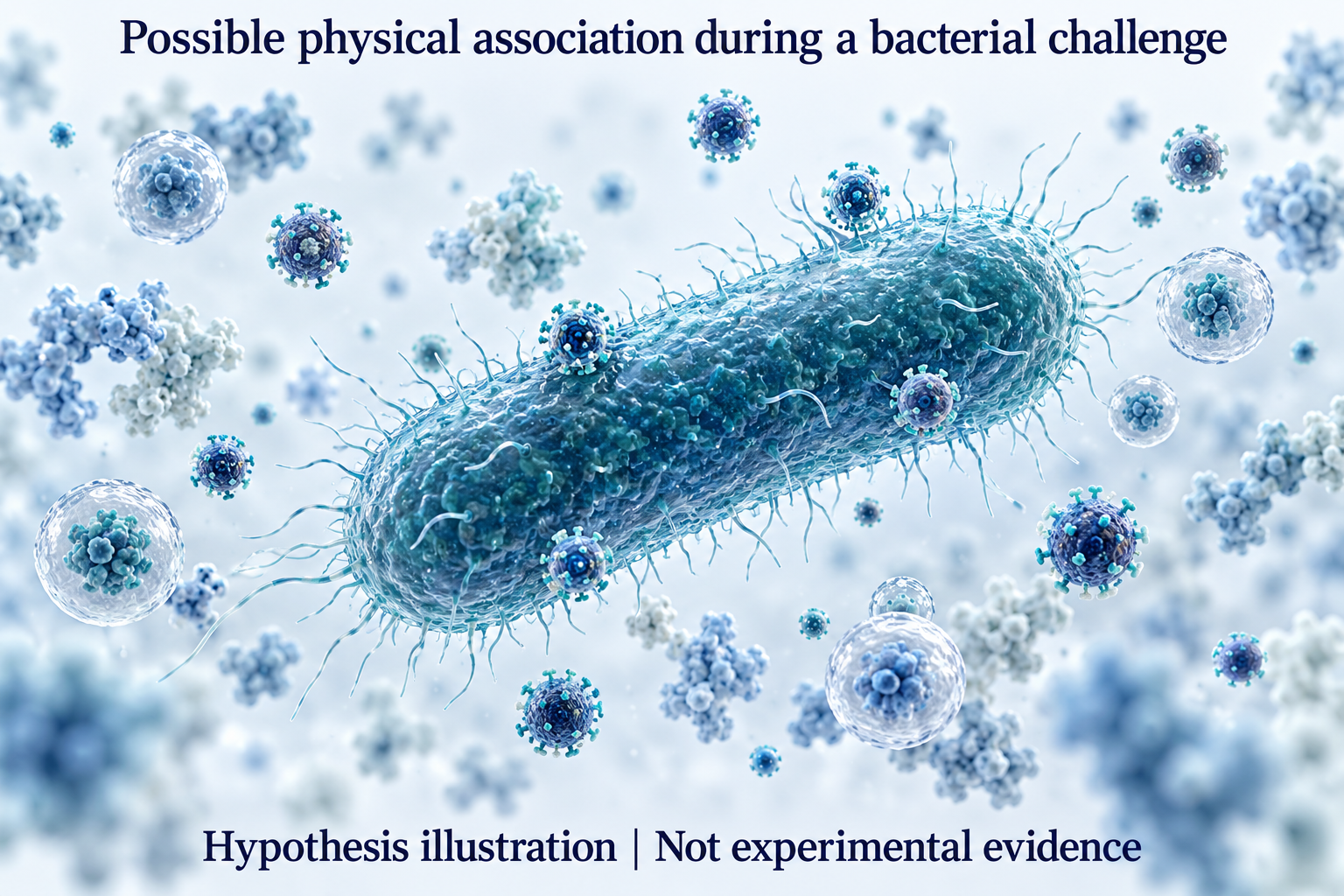
Figure 2 Hypothetical physical association of vector particles and other colloids with a bacterial surface. This illustration is not evidence of lentivirus binding to B. diminuta, and does not depict bacterial infection.
The evidence supports investigation of soft fouling
The rapid increase in resistance, incomplete throughput and contrast with the vector-free study are consistent with a fouling mechanism. The subsequently reported particle changes add evidence of altered behaviour in the challenge mixture.[11] These observations support investigation of soft fouling, but do not directly identify the deposited material or demonstrate a compressible gel on the membrane.
A membrane can become conditioned by material from the feed before a substantial deposit is apparent. Adsorption, pore restriction and surface accumulation may then contribute in different proportions as filtration proceeds.
Where deposited material is deformable and hydrated, it may form a compressible layer. In this context, 'soft fouling' describes a physical behaviour; 'gel-like' describes a proposed structure. Neither term establishes the chemical composition of the deposit or demonstrates that a gel has actually formed.
Research using model soft colloids has shown how cake compaction can reduce the spaces available for liquid flow and increase resistance.[4] This supports the general mechanism, but those model systems are not direct evidence of the same deposit in a lentiviral process.
If a compressible layer is present, increasing the driving pressure may compact it further, limiting the improvement in flow. At constant flow, additional resistance appears as a pressure rise; at constant pressure, it appears primarily as a decline in flow. The operating mode is therefore essential to interpretation.
The layers of a dual-membrane filter may contribute differently, but their individual behaviour needs evidence. The device configuration should be verified against the relevant manufacturer and study records. Published lentiviral filtration work found that some dual-membrane filters performed well, reinforcing the need to assess the particular feed and device.[5]
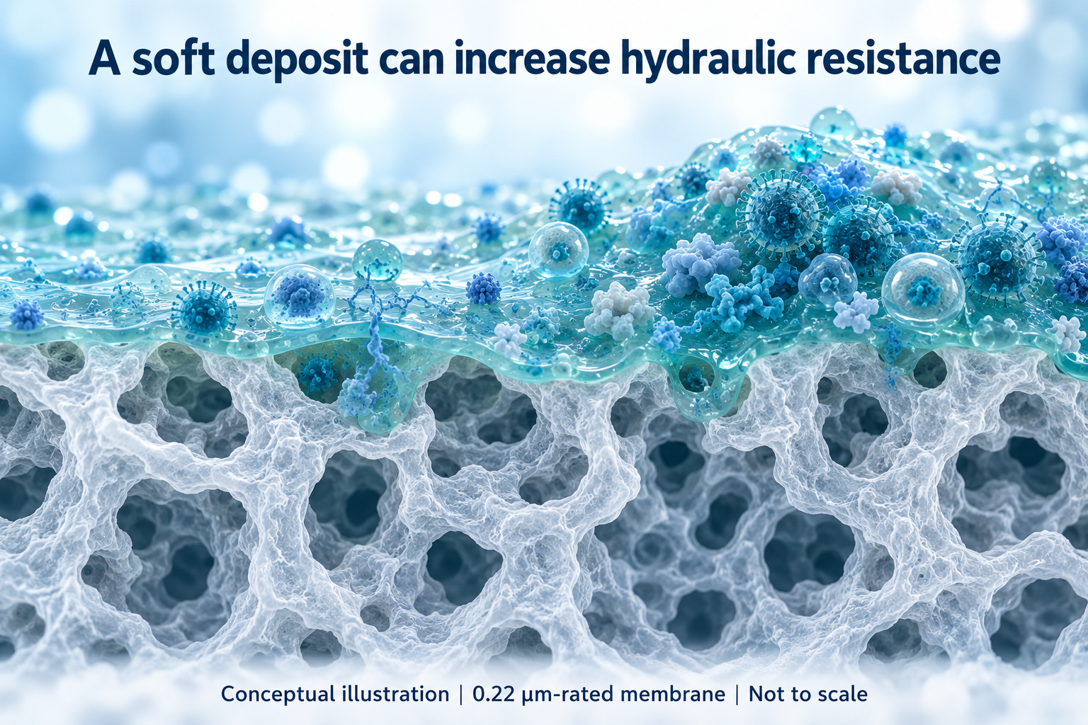
Figure 3 Hypothetical hydrated fouling layer over a 0.22 µm-rated membrane. Deposits may add resistance and restrict pore entrances. This is an artist's impression, not a micrograph or a confirmed deposit from a specific filtration study.
What the reported Videodrop experiment adds
A written follow-up account documents a completed Videodrop comparison of three preparations in tryptic soy broth (TSB): vector alone, B. diminuta alone and their mixture.[11] This adds qualitative experimental evidence to the earlier investigation proposal. The reported observations were:
Vector in TSB: the expected particle-size profile and particle number.
B. diminuta in TSB: a larger apparent particle size and the expected particle number.
B. diminuta plus vector in TSB: a pronounced reduction in measured particle number and an altered size profile.
The comparison supports a bacteria-associated change in the measurable particle population under the conditions examined. It is consistent with aggregation or physical association. However, particle sizing does not establish the identity of the altered population, and a lower measured count is not necessarily a loss of vector particles. Dilution corrections, mixing and hold times, matrix effects and the detection range need consideration.
The account is documentary evidence that the experiment and its outcome were reported; it does not supply the underlying size distributions, numerical concentrations, replicate results or direct imaging of a filter deposit. The strength and reproducibility of the effect therefore cannot be assessed independently from the material supplied.
The retained charts below are capability examples using polystyrene beads and extracellular vesicles. They are not the results of this three-condition experiment. Panels A–C show bead populations and mixed distributions; Panel D compares measured and theoretical extracellular-vesicle concentrations. The fitted slopes of approximately 0.45–0.47 also illustrate why dilution linearity should be distinguished from quantitative accuracy.
For the standard configuration described by the manufacturer, the quoted biological sizing range is 80–500 nm, with larger objects visible by microscopy up to 10 µm.[10] Movement outside the quantitative range or other detection effects could contribute to a lower count. Raw outputs, suitable controls and an orthogonal method would help distinguish these explanations.
The reported mixture behaviour gives a substantive reason to examine whether the challenge changes product filterability. A deliberately severe bacterial challenge differs from routine processing by design; the observation alone does not invalidate that challenge or establish that a surrogate will be representative.
Videodrop capability charts
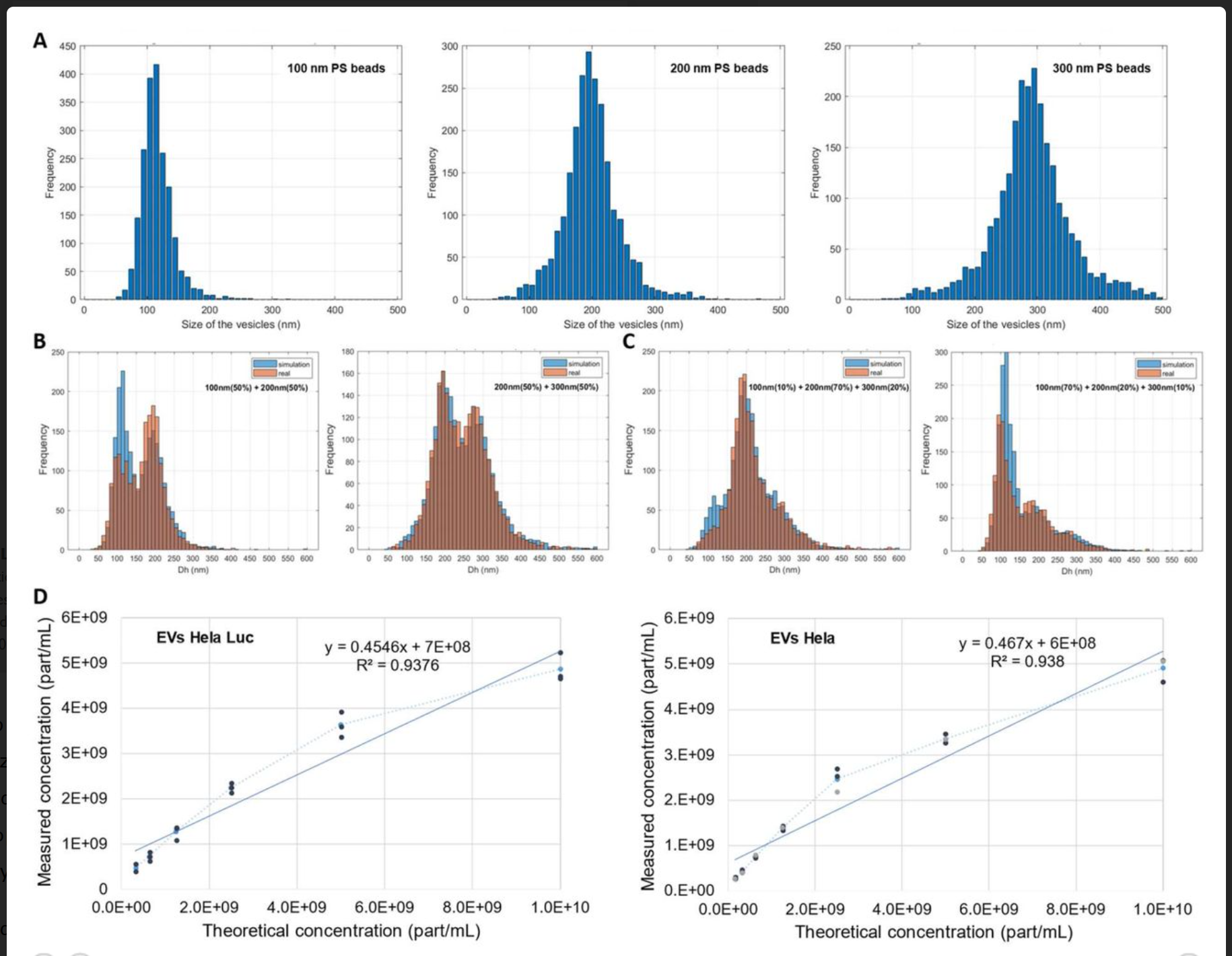
Figure 4 Videodrop capability charts retained from the technical review materials. A: nominal 100, 200 and 300 nm polystyrene beads. B and C: mixed bead populations with simulated and measured distributions. D: measured versus theoretical extracellular-vesicle concentrations. These charts do not show a lentiviral vector–bacteria mixture or a filter deposit. Scientific labels, axes, fits and plotted values are unchanged.
An immediate pressure rise has several possible causes
Rapid adsorption or deposition could contribute to high initial resistance. Equally, the assembly may contain trapped gas, an incompletely wetted membrane, a restricted connection or an unsuitable flow setting. Fluid viscosity, temperature and the preparation of the challenge suspension can also influence the result.
An upstream pressure reading should not automatically be reported as the pressure difference across the membrane. The locations of the sensors and any downstream backpressure need to be understood. Where two membrane layers are present, a total pressure measurement does not identify which layer contributes most resistance.
The pressure, flow, time and throughput records should be reviewed together from the point of start-up. Very low throughput at high pressure warrants an initial check of assembly and operating conditions before assigning a molecular explanation. Conditioning, cake formation and pore restriction may overlap, and their timing needs to be established for the system examined.
p24 and VCN require different interpretations
Differences between p24 recovery and a VCN-derived result can provide an important investigation signal, but the assays do not measure the same attribute.
A p24 assay measures capsid antigen within the capabilities of the method and its sample preparation. It does not independently establish infectivity or distinguish all intact particles from free or disrupted material. A direct vector-genome assay measures detectable sequences and, without suitable controls, does not establish an intact or functional vector.[6]
VCN usually refers to vector copy number measured in cells. Where a titre method includes cell transduction followed by PCR, the result reflects that biological and analytical workflow. Published methods use this approach to determine infectious titre and VCN in modified cells.[7] The term VCN alone should not be interpreted as a count of intact viral cores.
A higher p24 recovery than functional recovery may be consistent with loss of activity, differential retention or damage, but it does not identify which occurred. Assay variability, matrix effects, sampling, dilution and hold times also warrant consideration. Compare total recovered amounts, with the relevant volumes and dilution factors, rather than concentrations alone.
Orthogonal results are most useful when they help distinguish retention from loss of function and analytical effects. They should not be used to conclude that high pressure has disrupted the particles without additional evidence.
Build an investigation that can distinguish the explanations
A structured comparison can help establish when the additional resistance develops. Subject to the approved study design, relevant comparisons include buffer, unchallenged product, the challenge in an appropriate control vehicle and challenged product. An inoculum-vehicle control may help separate the effects of the added liquid from those of the organisms.
Use matched conditions and account for lot variability, membrane area and configuration, wetting, temperature, hold history, operating mode and loading per unit area. Volume alone may be an incomplete basis for comparison when vector and colloid concentrations differ.
Support the reported particle comparison with raw outputs, matched dilutions and reproducibility checks. Residual impurity measurements and deposit examination may help, although sizing alone cannot identify vector, vesicle, bacterium or aggregate. A dried deposit may differ substantially from a hydrated layer during operation.
The resulting explanation should account for the controls, the pressure and flow history, product recovery and microbiological results. More than one mechanism may contribute.
The validation strategy must remain scientifically justified
The initial review considered a size-matched polystyrene surrogate, a reduced bacterial challenge in vector-containing material, and a combination of those approaches.[9] The written follow-up proposed developing a surrogate rationale and protocol, with bead assessment for viability and size behaviour in the presence and absence of B. diminuta, followed by regulatory discussion before formal validation.[11] These were planned next steps; the supplied account does not demonstrate surrogate qualification, an executed alternative study or regulatory acceptance.
A 100 nm bead can be useful for investigating size-related effects, but matching size does not reproduce an enveloped vector’s surface chemistry, deformability or interaction with the membrane. A surrogate that removes the suspected interaction may also remove the very effect that needs to be assessed. Its relevance must therefore be demonstrated for the validation purpose.
Similarly, lowering the bacterial concentration does not automatically justify lowering the cumulative challenge per unit filter area. Concentration, total organisms delivered, exposure time and processed volume need separate consideration. Any alternative must retain an adequately justified challenge and remain applicable to the manufacturing conditions.
EU GMP Annex 1 calls for worst-case validation and, wherever possible, use of the product in bacterial retention testing. A surrogate requires justification when the product is unsuitable. Relevant parameters include conditioning, contact time, pressure, flow, volume and temperature.[8]
An alternative study design should address organism viability and recovery, the challenge delivered, integrity testing and its relationship to the product process. Passing a matrix-only study, or executing three surrogate runs with controls, does not automatically establish compliance for all product conditions.
The intended outcome is a study that can be completed and provides meaningful retention evidence within a defensible operating envelope. Acceptance of the rationale should be established prospectively through the appropriate quality and regulatory channels.
The practical conclusion
This case combines operational evidence of a filtration restriction with a documented qualitative report of altered particle behaviour after bacteria and vector were mixed. The findings support investigation of a challenge-associated interaction. Soft fouling remains a credible working explanation, while direct binding and the composition and compressibility of the proposed deposit remain to be established.
In my view, the strongest approach connects the pressure and recovery records with the raw particle measurements and appropriate controls, then uses that evidence to justify the retention study design. The written account advances the investigation. Its value is best preserved by distinguishing the reported result from the mechanism it may explain.
References
[1] Do Minh A et al. Characterization of Extracellular Vesicles Secreted in Lentiviral Producing HEK293SF Cell Cultures. Viruses. 2021;13:797.https://doi.org/10.3390/v13050797
[2] FDA. Sterile Drug Products Produced by Aseptic Processing — Current Good Manufacturing Practice. September 2004. Section IX B, Filtration Efficacy.https://www.fda.gov/media/71026/download
[3] Helling A et al. Investigation of microbial cell deformability by filter cake compressibility using ultrafiltration membranes. Colloids and Surfaces B Biointerfaces. 2020;185:110626.https://doi.org/10.1016/j.colsurfb.2019.110626
[4] Lüken A et al. Particle movements provoke avalanche-like compaction in soft colloid filter cakes. Scientific Reports. 2021;11:12836.https://doi.org/10.1038/s41598-021-92119-w
[5] Vogel JE et al. High yield sterile filtration process for highly concentrated lentiviral vectors. Biotechnology Journal. 2024;19:e2300348.https://doi.org/10.1002/biot.202300348
[6] Highly efficient concentration of lenti- and retroviral vector preparations by membrane adsorbers and ultrafiltration. BMC Biotechnology. 2011;11:55. Physical and biological titre comparisons.https://pmc.ncbi.nlm.nih.gov/articles/PMC3118112/
[7] Kandell J et al. Universal ddPCR-based assay for the determination of lentivirus infectious titer and lenti-modified cell vector copy number. Molecular Therapy Methods and Clinical Development. 2023;31:101120.https://doi.org/10.1016/j.omtm.2023.101120
[8] European Commission. EU GMP Annex 1 Manufacture of Sterile Medicinal Products. 2022 revision. Sections 8.81 and 8.83 to 8.87.https://health.ec.europa.eu/medicinal-products/eudralex/eudralex-volume-4_en
[9] Anonymised internal technical review of a sterilising-filter bacterial challenge programme. Reported observations and proposed follow-up activities, including the retained Videodrop capability charts. Underlying executed study reports and raw mixture-sizing data are not reproduced here.
[10] Corning. Nanoparticles Size and Concentration Characterization by Corning Videodrop. Application note CLS-AN-815. Quoted ranges apply to the configuration described in that document.https://www.corning.com/catalog/cls/documents/application-notes/CLS-AN-815.pdf
[11] Anonymised written follow-up communication reporting a completed Videodrop comparison of lentiviral vector, B. diminuta and their mixture in tryptic soy broth, together with proposed surrogate-development activities. Qualitative account; raw particle outputs and replicate data were not supplied.
Image notes
Figures 1 to 3 are AI-generated conceptual illustrations, not microscopy or experimental observations. Figure 4 retains the Videodrop capability charts from the technical review materials with their scientific content unchanged. It shows bead and extracellular-vesicle examples, not results from the vector–bacteria challenge mixture. Dates and identifying details of the internal programme have been removed; public reference publication dates and scientific chart labels are retained.